Fil:Wuchereria bancrofti LifeCycle.gif

Wuchereria_bancrofti_LifeCycle.gif(513 × 435 pikslar, filstorleik: 33 KB, MIME-type: image/gif)

Denne fila er frå Wikimedia Commons og kan verta nytta av andre prosjekt. Skildringa frå filskildringssida der er vist nedanfor.


Følgjande er henta frå filomtalen åt denne fila på Wikimedia Commons:


Skildring

Wuchereria bancrofti

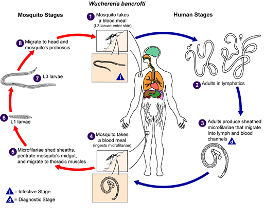
Life cycle of Wuchereria bancrofti

Different species of the following genera of mosquitoes are vectors of W. bancrofti filariasis depending on geographical distribution. Among them are: Culex (C. annulirostris, C. bitaeniorhynchus, C. quinquefasciatus, and C. pipiens); Anopheles (A. arabinensis, A. bancroftii, A. farauti, A. funestus, A. gambiae, A. koliensis, A. melas, A. merus, A. punctulatus and A. wellcomei); Aedes (A. aegypti, A. aquasalis, A. bellator, A. cooki, A. darlingi, A. kochi, A. polynesiensis, A. pseudoscutellaris, A. rotumae, A. scapularis, and A. vigilax); Mansonia (M. pseudotitillans, M. uniformis); Coquillettidia (C. juxtamansonia). During a blood meal, an infected mosquito introduces third-stage filarial larvae onto the skin of the human host, where they penetrate into the bite wound. They develop in adults that commonly reside in the lymphatics. The female worms measure 80 to 100 mm in length and 0.24 to 0.30 mm in diameter, while the males measure about 40 mm by .1 mm. Adults produce microfilariae measuring 244 to 296 μm by 7.5 to 10 μm, which are sheathed and have nocturnal periodicity, except the South Pacific microfilariae which have the absence of marked periodicity. The microfilariae migrate into lymph and blood channels moving actively through lymph and blood. A mosquito ingests the microfilariae during a blood meal. After ingestion, the microfilariae lose their sheaths and some of them work their way through the wall of the proventriculus and cardiac portion of the mosquito's midgut and reach the thoracic muscles. There the microfilariae develop into first-stage larvae and subsequently into third-stage infective larvae. The third-stage infective larvae migrate through the hemocoel to the mosquito's prosbocis and can infect another human when the mosquito takes a blood meal.
Kjelde http://www.dpd.cdc.gov/dpdx/images/ParasiteImages/A-F/Filariasis/W_bancrofti_LifeCycle.gif
Opphavsperson
Løyve
(Gjenbruk av denne fila)
Public domain
This image is a work of the Centers for Disease Control and Prevention, part of the United States Department of Health and Human Services, taken or made as part of an employee's official duties. As a work of the U.S. federal government, the image is in the public domain.

eesti  Deutsch  čeština  español  português  English  français  Nederlands  polski  slovenščina  suomi  македонски  українська  日本語  中文(简体)‎  中文(繁體)‎  العربية  +/−

Bilettekstar

Skriv inn ei line med tekst som skildrar fila

Element som er med i denne fila

motiv

image/gif

Filhistorikk

Klikk på dato/klokkeslett for å sjå fila slik ho var på det tidspunktet.

Dato/klokkeslettMiniatyrbileteOppløysingBrukarKommentar
gjeldande5. november 2008 kl. 16:15Miniatyrbilete av versjonen frå 5. november 2008 kl. 16:15513 × 435 (33 KB)LycaonWatermark removed
14. mai 2006 kl. 23:02Miniatyrbilete av versjonen frå 14. mai 2006 kl. 23:02513 × 435 (36 KB)Patho{{Information| |Description=Filariasis [Brugia malayi] [Brugia timori] [Loa loa] [Mansonella ozzardi] [Mansonella perstans] [Mansonella streptocerca] [Onchocerca volvulus] [Wuchereria bancrofti] Life cycle of Wuchereria bancrofti Different species

Den følgjande sida bruker denne fila:

Global filbruk

Desse andre wikiane nyttar fila: